Hip Procedures
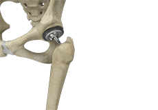
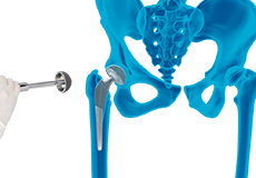
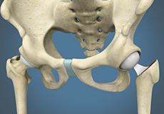
Total Hip Replacement
Total hip replacement is a surgical procedure in which the damaged cartilage and bone are removed from the hip joint and replaced with artificial components. The main indication for total hip replacement is arthritis.
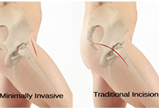
Minimally Invasive Total Hip Replacement
Minimally invasive total hip replacement is a surgical procedure performed through one or two small incisions rather than the single long incision of 10–12-inches as in the traditional approach.
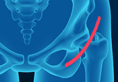
Anterior Hip Replacement
Anterior hip replacement surgery is performed under general anesthesia or regional anesthesia. You will lie down on your back, on a special operating table that enables your surgeon to perform the surgery from the front of the hip. Your surgeon may use fluoroscopic imaging during the surgery to ensure the accuracy of component positioning and to minimize leg length inequality.
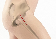
Outpatient Anterior Approach Hip Replacement
Outpatient anterior approach hip replacement refers to surgery accessed from in front of the hip in an outpatient setting. It is a minimally invasive procedure that has been developed to cause less muscle damage, faster recovery, and less disruption in a patient’s life.
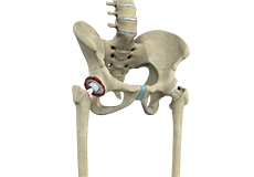
Robotic Total Hip Replacement
Robotic total hip replacement is a minimally invasive procedure where your surgeon is assisted by a robotic system to perform a total hip replacement surgery.
Revision Hip Replacement
Revision hip replacement is a complex surgical procedure in which all or part of a previously implanted hip joint is replaced with a new artificial hip joint. Total hip replacement surgery is an option to relieve severe arthritis pain that limits your daily activities.
Hip Trauma Reconstruction
Hip trauma is an injury in the hip due to the impact caused by incidents such as a car accident or a hard fall. The injury can be a bone break or dislocation or both. Hip trauma reconstruction is the process of rebuilding and restoring the hip joint.
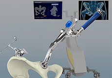
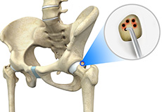
Computer-assisted Hip Replacement
Computer-assisted hip replacement is an image-guided, minimally invasive surgical procedure to replace your diseased or damaged hip with an artificial device using the assistance of computer software. The system creates and displays images and provides information that aids your surgeon at various stages of the procedure to improve accuracy and results.
Hip Hemiarthroplasty
Hip hemiarthroplasty is a surgical technique employed to treat hip fractures. In this procedure, only one half (ball section) of the hip joint is substituted by a metal prosthesis.
Hip Fracture Surgery
Hip fractures involve a break that occurs near the hip in the upper part of the femur or thigh bone. The thigh bone has two bony processes on the upper part - the greater and lesser trochanters. The lesser trochanter projects from the base of the femoral neck on the back of the thigh bone. Hip fractures can occur either due to a break in the femoral neck, in the area between the greater and lesser trochanter or below the lesser trochanter.
Hip Microfracture
Hip Microfracture is a marrow-stimulating technique that creates a network of small holes in the bone below the hip cartilage (subchondral bone). The goal of the procedure is to increase the blood supply to stimulate cartilage growth.
Physical Therapy for Hip
Physical therapy is an exercise program that helps you to improve movement, relieve pain, encourage blood flow for faster healing, and restore your physical function and fitness level. The main aim of physical therapy is to make your daily activities, such as walking, getting in and out of bed and climbing stairs, easier.
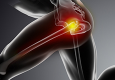
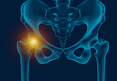
Hip Conditions
Hip Flexor Pain
Hip flexor pain is a distressing feeling or discomfort noted in the hip and/or groin region that can make everyday activities, such as going up and down the stairs or lifting your leg to tie a shoe extremely difficult and painful and can severely limit your activity and mobility.
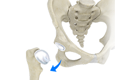
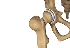
Hip Dislocation
The hip joint is a “ball and socket” joint. The “ball” is the head of the femur or thighbone, and the “socket” is the cup-shaped acetabulum. The joint is surrounded by muscles, ligaments, and tendons that support and hold the bones of the joint in place. Hip dislocation occurs when the head of the femur moves out of the socket.
Hip Fracture
A hip fracture is a break that occurs near the hip in the upper part of the femur or thighbone. The thighbone has two bony processes on the upper part - the greater and lesser trochanters. The lesser trochanter projects from the base of the femoral neck on the back of the thighbone.
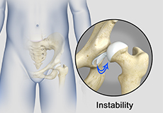
Hip Instability
Injury or damage to these structures can lead to a condition called hip instability when the joint becomes unstable.
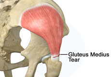
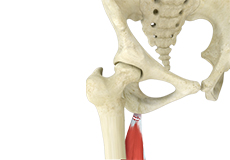
Gluteus Medius Tear
A gluteus medius tear is the partial or complete rupture of the gluteus medius muscle due to severe muscle strain. Gluteus medius tears often occur at the tendinous attachment to the greater trochanter of the femur bone.
Hip Labral Tear
A hip labral tear is an injury to the labrum, the cartilage that surrounds the outside rim of your hip joint socket.
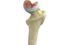
Hip Osteonecrosis
Hip osteonecrosis occurs due to disruption of the blood supply to the highest part of the thigh bone (femoral head). Due to lack of nourishment, the bone tissue of the femoral head dies and gradually collapses, which may further lead to degeneration of the underlying cartilage.
Hip Bursitis
Hip bursitis is a painful condition caused by the inflammation of a bursa in the hip. Bursae are fluid-filled sacs present in the joints between bone and soft tissue to reduce friction and provide cushioning during movement.
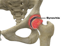
Hip Synovitis
Hip synovitis, also called transient hip synovitis or toxic synovitis, is a condition characterized by inflammation of the synovial tissues that surround the hip joint. It is the most common cause for sudden hip pain that occurs in young children between the age of 2 and 9. It affects boys more commonly than girls and is most of the time limited to only one side of the hip.
Iliopsoas Tendonitis
Iliopsoas tendonitis also referred to as snapping hip syndrome, is an inflammation of the iliopsoas tendon or the surrounding area. The iliopsoas is the hip flexor tendon located over the front of the hip socket. The term snapping hip describes the sound made, a snap or click, that occurs with certain hip movements including flexion, extension, and rotation of the hip.
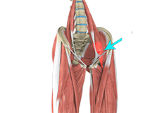
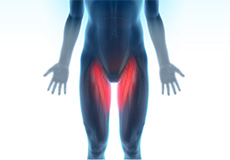
Hip Adductor Injuries
Hip adductors are the group of muscles on the inner side of your thigh that enable adduction or the ability to bring the thighs together.
Hip Ligament Injuries
Injuries to the hip ligaments are commonly called a hip sprain and can range from minor tears of the ligaments to more serious injuries involving the hip muscles, tendons or bone.
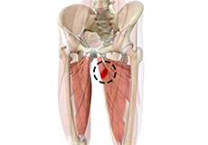
Groin Injuries in Athletes
Groin injuries are injuries sustained by athletes during sports activity. Groin injuries comprise about 2 to 5 percent of all sports injuries. The most common kind of groin injury is a groin strain or a pulled groin muscle. Typical groin injuries vary based on athlete’s gender, age, and sports, with most common groin injuries occurring in certain sports such as football with a prevalence rate as high as 12-16%.
Hamstring Injuries
The hamstring is a group of three muscles that run along the back of the thigh from the hip to the knee. Hamstring injuries occur when these muscles are strained or pulled. They are common in dancers and athletes of all sorts including runners and those who play football, soccer, basketball, tennis, etc.
Hip Anatomy
Hip Joint
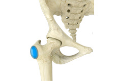
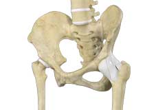
The hip joint is the largest weight-bearing joint in the human body. It is also referred to as a ball and socket joint and is surrounded by muscles, ligaments, and tendons. The thigh bone or femur and the pelvis join to form the hip joint.
Any injury or disease of the hip will adversely affect the joint's range of motion and ability to bear weight.
The hip joint is made up of the following:
- Bones and joints
- Ligaments of the joint capsule
- Muscles and tendons
- Nerves and blood vessels that supply the bones and muscles of the hip
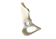
Bones and Joints
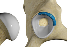
The hip joint is the junction where the hip joins the leg to the trunk of the body. It is comprised of two bones: the thigh bone or femur and the pelvis which is made up of three bones called ilium, ischium, and pubis. The ball of the hip joint is made by the femoral head while the socket is formed by the acetabulum. The Acetabulum is a deep, circular socket formed on the outer edge of the pelvis by the union of three bones: ilium, ischium, and pubis. The lower part of the ilium is attached by the pubis while the ischium is considerably behind the pubis. The stability of the hip is provided by the joint capsule or acetabulum and the muscles and ligaments which surround and support the hip joint.
The head of the femur rotates and glides within the acetabulum. A fibrocartilagenous lining called the labrum is attached to the acetabulum and further increases the depth of the socket.
The femur or thigh bone is one of the longest bones in the human body. The upper part of the thigh bone consists of the femoral head, femoral neck, and greater and lesser trochanters. The head of the femur joins the pelvis (acetabulum) to form the hip joint. Next, to the femoral neck, there are two protrusions known as greater and lesser trochanters which serve as sites of muscle attachment.
Articular cartilage is the thin, tough, flexible, and slippery surface lubricated by synovial fluid that covers the weight-bearing bones of the body. It enables smooth movements of the bones and reduces friction.
Ligaments
Ligaments are fibrous structures that connect bones to other bones. The hip joint is encircled with ligaments to provide stability to the hip by forming a dense and fibrous structure around the joint capsule. The ligaments adjoining the hip joint include:
- Iliofemoral ligament: This is a Y-shaped ligament that connects the pelvis to the femoral head at the front of the joint. It helps in limiting the over-extension of the hip.
- Pubofemoral ligament: This is a triangular shaped ligament that extends between the upper portion of the pubis and the iliofemoral ligament. It attaches the pubis to the femoral head.
- Ischiofemoral ligament: This is a group of strong fibers that arise from the ischium behind the acetabulum and merge with the fibers of the joint capsule.
- Ligamentum teres: This is a small ligament that extends from the tip of the femoral head to the acetabulum. Although it has no role in hip movement, it does have a small artery within that supplies blood to a part of the femoral head.
- Acetabular labrum: The labrum is a fibrous cartilage ring which lines the acetabular socket. It deepens the cavity, increasing the stability and strength of the hip joint.
Muscles and Tendons
A long tendon called the iliotibial band runs along the femur from the hip to the knee and serves as an attachment site for several hip muscles including the following:
- Gluteals: These are the muscles that form the buttocks. There are three muscles (gluteus minimus, gluteus maximus, and gluteus medius) that attach to the back of the pelvis and insert into the greater trochanter of the femur.?
- Adductors: These muscles are located in the thigh which helps in adduction, the action of pulling the leg back towards the midline.?
- Iliopsoas: This muscle is located in front of the hip joint and provides flexion. It is a deep muscle that originates from the lower back and pelvis and extends up to the inside surface of the upper part of the femur.?
- Rectus femoris: This is the largest band of muscles located in front of the thigh. They also are hip flexors.?
- Hamstring muscles: These begin at the bottom of the pelvis and run down the back of the thigh. Because they cross the back of the hip joint, they help in extension of the hip by pulling it backward.
Nerves and Arteries
Nerves of the hip transfer signals from the brain to the muscles to aid in hip movement. They also carry the sensory signals such as touch, pain, and temperature back to the brain.
The main nerves in the hip region include the femoral nerve in the front of the femur and the sciatic nerve at the back. The hip is also supplied by a smaller nerve known as the obturator nerve.
In addition to these nerves, there are blood vessels that supply blood to the lower limbs. The femoral artery, one of the largest arteries in the body, arises deep in the pelvis and can be felt in front of the upper thigh.
Hip Movements
All of the anatomical parts of the hip work together to enable various hip movements. Hip movements include flexion, extension, abduction, adduction, circumduction, and hip rotation.